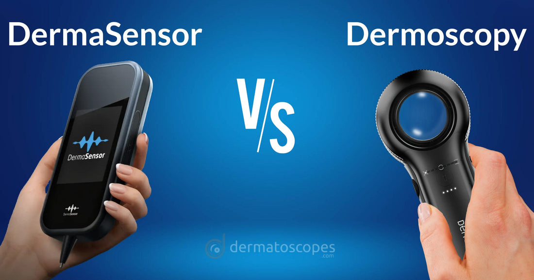
As a clinician whose responsibilities include that of skin cancer screening, you may have heard about the new DermaSensor device.
Is it worth adding to your practice? Or is “dermoscopy” still your best choice?
The Importance of Early Detection
Skin cancer is far and away the most common type of cancer in the US.1 In fact, it accounts for nearly half of all cancers.
Although nearly 20 Americans die every day from skin cancer (primarily melanoma), statistically most skin cancers aren’t “deadly.” Even those which aren’t life-threatening, though, still carry the risk of significant disfigurement, pain, and loss of function – especially if not treated early.
The Role of Technology in Diagnosis
Technological advancements are transforming how we diagnose skin cancer. Traditional methods like visual inspection are now supplemented with sophisticated tools.
Two notable tools are DermaSensor and dermoscopy. Each has its advantages and disadvantages.
DermaSensor
-
Pros
- Not dependent on knowledge of skin cancer screening criteria
- High sensitivity
- Rivals that of screening performed by experienced dermatologists
-
Cons
- Low specificity
- A fair number of lesions “flagged” by the device will ultimately prove to be benign
- Additional expense (subscription-based plan)
- ~$200/month (for up to 5 screenings per month), or…
- ~$400/month (for unlimited screenings)
- Not practical for general lesion screening due to time/steps involved
- Requires WiFi connection to perform
- Generally not an issue, but worth considering for rural/remote screenings
- Low specificity
Dermoscopy
-
Pros
- Faster than DermaSensor
- Trained dermoscopists generally need less than 10 seconds per lesion
- Less costly
- Professional-level dermatoscopes currently average ~$1500/ea
- Both sensitive and specific
- Images obtained via dermoscopy also facilitate documentation as well as collaboration (when needed)
- Faster than DermaSensor
-
Cons
- Dependent on user’s knowledge and expertise
DermaSensor: Behind the Technology
DermaSensor devices use a technology called "Elastic Scattering Spectroscopy" (ESS) to assess skin lesions for potential malignancies. Here's how it works:
Light Scattering
ESS involves shining specific wavelengths of light onto the skin and analyzing the light scattered back. Skin tissue, which includes cells, collagen fibers, and other microscopic structures, scatters light in characteristic ways. The pattern of scattered light provides information about tissue composition, structure, and cellular changes.
Spectral Analysis
The device captures the unique spectral "fingerprint" of the light scattered from the skin tissue. Variations in the spectrum reflect differences in the tissue’s cellular and structural makeup, which can help identify abnormal changes consistent with malignant or benign lesions.
Data Interpretation with Machine Learning
DermaSensor devices incorporate machine learning algorithms trained on extensive datasets of skin lesion spectra. These algorithms compare the captured spectral data from a patient’s skin lesion to a large library of spectra from known benign and malignant lesions to determine the probability that a lesion may be cancerous.
Dermoscopy
Dermoscopy is a non-invasive diagnostic technique that enhances visualization of skin lesions, helping clinicians differentiate between benign and malignant lesions.
Magnified Imaging
Dermoscopy uses a handheld device called a dermatoscope, which combines magnification (typically 10x or more) and a polarized or non-polarized light source to illuminate the skin lesion. The device allows clinicians to see details not visible to the naked eye.
Surface Reflection Reduction
Non-polarized dermoscopy requires contact with the skin, often with a gel or oil to reduce surface reflection, allowing light to penetrate the outer layers of skin. Polarized dermoscopy doesn’t require contact, as it uses cross-polarized light to minimize surface reflections naturally.
Visualization of Dermoscopic Structures
By looking beneath the stratum corneum (the outermost layer of the skin), dermoscopy reveals specific structures, patterns, and colors within the lesion that are diagnostic for various skin conditions. Common features include pigment networks, vascular structures, and other patterns indicative of melanocytic (melanoma-prone) and non-melanocytic lesions.
Pattern Analysis and Diagnostic Algorithms
Clinicians use dermoscopic criteria and pattern analysis to identify lesion characteristics associated with malignancies, like melanomas, or benign conditions, like seborrheic keratosis. They may apply diagnostic algorithms, such as the "ABCDE" criteria or "Chaos and Clues," to assess risk.
The 'Ideal' Scenarios for Each
DermaSensor
The real advantage of DermaSensor is its ability to identify high-risk lesions which could have been missed due to the screener’s lack of experience, training, and/or expertise.
While providers who utilize DermaSensor for decision-making support are likely to “catch” melanomas at a rate equivalent to that of experienced dermatologists, they’re also more likely to biopsy lesions which ultimately prove to be benign. Remember – high sensitivity, low specificity.
Dermoscopy
Dermoscopy still remains the “gold standard” of skin cancer screening, but its not without its faults. However, dermoscopy's advantage is highly dependent on user training and expertise.
In fact, a study published in the British Journal of Dermatology compared the diagnostic performance of experienced dermatologists with that of clinicians with minimal dermoscopy training.2 The results showed that those with minimal dermoscopy training actually had lower sensitivity (69%) in detecting melanomas compared to the experienced dermatologist (92%). This suggests that without sufficient training, the use of dermoscopy could potentially impair diagnostic accuracy.
On the other hand, dermoscopy performed with training has been shown to increase sensitivity by up to 25% and specificity by up to 10% (compared to naked eye examination).3
While this emphasis on proficiency may sound overwhelming, Binder et al showed dermoscopy improved diagnostic accuracy after 9 hours of training.4
The Flawed Premise
To a degree, the “DermaSensor vs. Dermoscopy” debate is based on a flawed premise which suggests that the decision itself is binary in nature. Instead, “Dermoscopy (Alone) vs. DermaSensor + Dermoscopy” would actually make for a more realistic argument.
"Instead, 'Dermoscopy (Alone) vs. DermaSensor + Dermoscopy' would actually make for a more realistic argument."
The truth is that most clinicians who are truly active in performing skin cancer screenings will at least incorporate dermoscopy into their practice. Unless time is of no significance, it’s simply not practical to use devices like DermaSensor on several or more lesions at every skin exam.
Therefore, the real question is…
Who Should 'Add' the DermaSensor to Their Skin Cancer Screening Protocol?
Any clinician who regularly performs skin cancer screenings and is unable (or unwilling) to acquire the knowledge necessary to proficiently perform dermoscopy will need a plan for managing patients whose lesions call for additional scrutiny.
By far, the most common choice in these situations would involve referral to a dermatologist. However, sometimes that’s easier said than done. In many parts of the country, patients (including those referred by their PCP) need to wait months before being seen by the dermatologist.
To be clear, situations in which patients have a particularly concerning lesion and barriers are present which may prevent the timely referral to a dermatologist, DermaSensor stands to make the greatest impact.
At the very least, if the patient’s PCP can screen these lesions with the DermaSensor and receive a negative result, the reassurance it provides will prove invaluable.
On the other hand, when the PCP’s DermaSensor device confirms their concerns, they'll be faced with two options. The first would be that the results could serve as added motivation for performing the biopsy themselves, thereby ensuring immediate intervention. Alternatively, if the PCP still chooses to refer the patient to a dermatologist, we need to consider what effect a preliminary 'DermaSensor screening' result may have on the referral dynamics.
An unspoken truth is that, for many dermatologists, their trust in the legitimacy of a referral can affect whether accommodations are made to squeeze patients into their already-packed schedules. Unfortunately, dermatologists have grown accustomed to receiving "urgent referrals" for lesions like seborrheic keratoses which, given their expertise, they quickly recognize as not being of any cause for concern.
But what about referrals which are accompanied by a "positive" DermaSensor result?
It stands to reason that dermatologists may treat such referrals with a greater sense of urgency. Only time will tell if that proves to be the case.
REFERENCES:
1. “Basal & Squamous Cell Skin Cancer Statistics.” Basal & Squamous Cell Skin Cancer Statistics | American Cancer Society, www.cancer.org/cancer/types/basal-and-squamous-cell-skin-cancer/about/key-statistics.html. Accessed 12 Nov. 2024.
2. British Journal of Dermatology, Volume 147, Issue 3, 1 September 2002, Pages 481–486, https://doi.org/10.1046/j.1365-2133.2002.04978.x
3. Tognetti, L. et al. (2020). Dermoscopy: Fundamentals and Technology Advances. In: Fimiani, M., Rubegni, P., Cinotti, E. (eds) Technology in Practical Dermatology. Springer, Cham. https://doi.org/10.1007/978-3-030-45351-0_1.
4. Binder M, Puespoeck-Schwarz M, Steiner A, et al. Epiluminescence microscopy of small pigmented skin lesions: Short-term formal training improves the diagnostic performance of dermatologists. J Am Acad Dermatol. 1997;36(2):197–202. doi: 10.1016/s0190-9622(97)70280-9.

.png?v=1667978305)
